Interleukins
Interleukins (1B, 6 & 10)
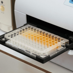
EGP250.00 / SampleIL-6 (Interleukin 6) ELISA Assay
Interleukin 6 (IL-6) is a multifunctional 26 kD protein originally discovered in the medium of RNA-stimulated fibroblastoid cells. IL-6 appears to be directly involved in the responses that occur after infection and cellular injury, and it may prove to be important as IL-1 and TNF-a in regulating the acute phase response. Primarily produced at sites of acute and chronic inflammation, IL-6 is secreted into the serum and induces a transcriptional inflammatory response through interleukin 6 receptor, alpha. The functioning of IL-6 is implicated in a wide variety of inflammation-associated disease states including diabetes mellitus and systemic juvenile rheumatoid arthritis.
Assay Principle
ELISA plates are pre-coated with an antibody specific to Rat IL-6. After adding samples/standards, a biotinylated detection antibody specific for Rat IL-6 and Avidin-Horseradish Peroxidase (HRP) conjugate are added successively to each well and incubated. The optical density of IL-6 conjugated with the biotinylated detection antibody is measured spectrophotometrically at a wavelength of 450 nm. The OD value is proportional to the concentration of Rat IL-6.
IL-1ꞵ (Interleukin 1 Beta) ELISA Assay
Interleukin-1 beta (IL-1 beta) is a proinflammatory cytokine expressed by monocytes, macrophages, and dendritic cells. IL-1 beta is synthesized in response to inflammatory stimuli as a 31 kDa inactive pro-form that accumulates in the cytosol. Cleavage of pro-IL-1 beta into the active 17 kDa protein requires the activation of inflammasomes, which are multi-protein complexes that respond to pathogens, stress conditions, and other danger signals. Inflammasome activation triggers the processing of the caspase-1 precursor into its active form, which in turn cleaves pro-IL-1 beta. IL-1 beta lacks a signal sequence peptide for classical ER/Golgi pathway and is secreted alongside caspase-1 via an alternate and incompletely understood mechanism. IL-1 beta play an important role in innate host defense by triggering the production of other proinflammatory cytokines in target cells and initiating acute-phase responses to infection and injury. Elevated levels of IL-1 beta have been associated with many chronic inflammatory conditions IL-1 beta neutralizing antibodies potential therapeutic value.
Assay Principle
The Rat IL-1β solid-phase sandwich ELISA (enzyme-linked immunosorbent assay) is designed to measure the amount of the target bound between a matched antibody pair. A target-specific antibody has been pre-coated in the wells of the supplied microplate. Samples, standards, or controls are then added into these wells and bind to the immobilized (capture) antibody. The sandwich is formed by the addition of the second (detector) antibody, a substrate solution is added that reacts with the enzyme-antibody-target complex to produce measurable signal. The optical density (OD) is measured spectrophotometrically at a wavelength of 450 nm
IL-10 (Interleukin 10) ELISA Assay
IL-10 (Interleukin-10) is a pleiotropic cytokine with important immunoregulatory functions. Its actions influence activities of many of the cell-types in the immune system. It is a cytokine with potent anti-inflammatory properties, repressing the expression of inflammatory cytokines such as TNF-Alpha (Tumor Necrosis Factor-Alpha), IL-6 (Interleukin-6) and IL-1 (Interleukin-1) by activated macrophages.
Assay Principal
The micro ELISA plate is pre-coated with an antibody specific to Rat IL-10. Standards/samples are added to the micro ELISA plate wells and combined with the specific antibody. Then a biotinylated detection antibody specific for Rat IL-10 and Avidin-Horseradish Peroxidase (HRP) conjugate are added successively to each micro plate well and incubated. Free components are washed awayl. Only those wells that contain Rat IL-10, biotinylated detection antibody and Avidin-HRP conjugate will appear blue in color. The enzyme-substrate reaction is terminated by the addition of stop solution and the color turns yellow. The optical density is measured spectrophotometrically at a wavelength of 450 nm. The OD value is proportional to the concentration of Rat IL-10.